Abstract
Introduction:
Detection of minimal residual disease (MRD) is one of the strongest predictors of outcome in multiple myeloma (MM). Until recently, the most commonly available method to detect MRD in clinical practice has been high sensitivity flow cytometry (FC) which can detect MRD with at 10 -5 sensitivity. In recent years, next-generation sequencing (NGS) has become a viable method to assess the MRD in MM patients with a 10 -6 sensitivity. NGS appears to have some advantages over HC-FC by circumventing subjectivity of analysis. However, real-world comparison between these two methodologies in the literature is limited and is important to inform daily hematopathology and oncology ordering practices.
Methods:
We retrospectively identified all cases of MM with NGS MRD data from bone marrow specimens at the Moffitt Cancer Center and collated corresponding flow MRD data and clinical data (OS, patient demographics) electronically and via chart review.
10-color flow cytometry was performed on a Gallios System and analyzed on Kaluza (Beckman Coulter, IN). Two million events were collected on all cells. Validated lower limit of detection was at least 0.01%. Antibodies included CD28, CD81, CD56, CD138, CD319, CD20, CD19, CD117, CD38, CD45, CD27, CD200 (BD, Biolegend, Beckman Coulter).
clonoSEQ ® (Adaptive Biotechnologies, Seattle, WA) testing was performed which uses multiplex polymerase chain reaction (PCR) and NGS to identify, characterize, and monitor clonotypes of immunoglobulin (Ig) IgH (V-J), IgH (D-J), IgK, and IgL receptor gene sequences, and translocated BCL1/IgH (J) and BCL2/IgH (J) sequences
Statistical analysis was performed by Spearman correlation coefficient and Kaplan-Meier analysis.
Results:
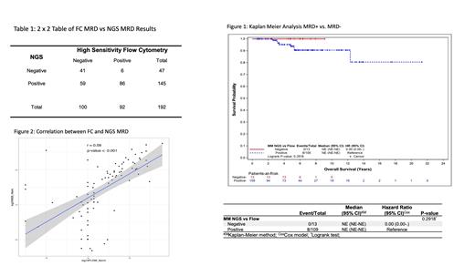
192 samples from 122 unique patients were identified that had both NGS and FC data performed on the same sample. FC+ values ranged from 1x10 -7 to 0.39. NGS+ values ranged from 2.3 x 10 -7 to 0.15. Spearman correlation coefficient showed moderate concordance between NGS and FC at r=0.67 (p<0.001). Six samples were positive by FC (mean tumor burden (MTB)= 0.0007) but missed by NGS; whereas 59 samples were positive by NGS (MTB= 0.002) but missed by flow cytometry. Two cases by FC were equivocal and these were both definitively designated as MRD+ by NGS. Overall survival was worse for MRD+ (by NGS or FC) vs MRD(-) (Figure 1).
Conclusion:
Our study confirms the importance of MRD detection in MM and shows the robust utility of NGS for MRD detection in routine hematopathology practice. While both FC and NGS are complementary given that each can potentially detect MRD missed by another method, the data supports the increased sensitivity of NGS over FC.
Nishihori: Novartis: Research Funding; Karyopharm: Research Funding. Hussaini: Stemeline Therapeutics: Honoraria.


This feature is available to Subscribers Only
Sign In or Create an Account Close Modal